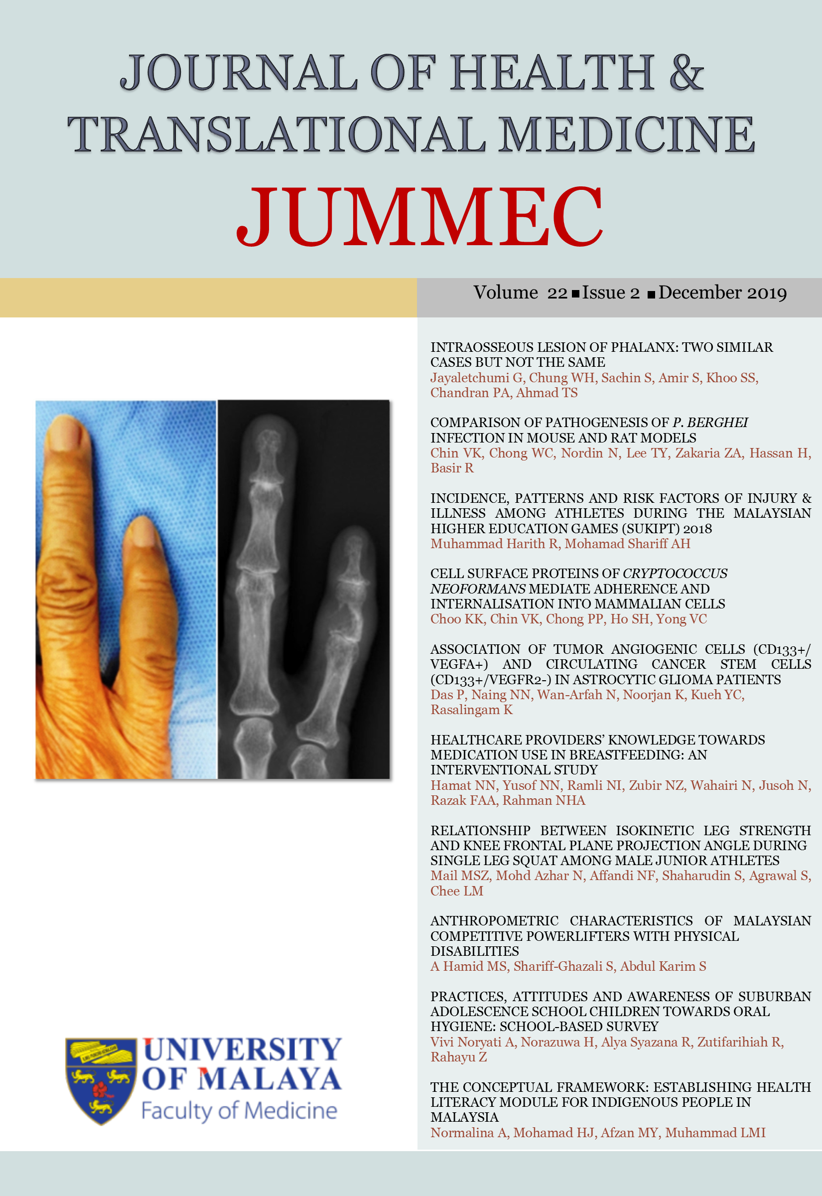
INTRAOSSEOUS LESION OF PHALANX: TWO SIMILAR CASES BUT NOT THE SAME
Received 2018-10-16; Accepted 2019-04-25; Published 2019-07-29
DOI:
https://doi.org/10.22452/jummec.vol22no2.1Keywords:
Intraosseous lesions, intraosseous gout, Epithelioid hemangioma, phalanxAbstract
Intraosseous lesions at phalanges are rare. They frequently present with pain and swelling. Fortunately, majority of the lesions are benign. However, some lesions are destructive and early interventions are required. We report two cases of similar presentations of swelling and discomfort at little finger for six months. The lytic lesions involved the whole middle phalanx with cortical breach sparing joints. Diagnosis was impossible with imaging alone. Bone biopsy was performed early to plan definitive treatment and surgery. Patient 1 was diagnosed for intraosseous gout whereas Patient 2 for epithelioid hemangioma. Both were benign destructive bone lesions. Thus, we counselled for curettage of lesion, bone grafting and spanning external fixation in view of extensive lesion. Patient 1 had defaulted treatment. Patient 2 had uneventful surgery. She regained her grip strength. In two years follow up, there was no evidence of infection, recurrence or malignant transformation.
Downloads
Downloads
Published
Issue
Section
License
All authors agree that the article, if editorially accepted for publication, shall be licensed under the Creative Commons Attribution License 4.0 to allow others to freely access, copy and use research provided the author is correctly attributed, unless otherwise stated. All articles are available online without charge or other barriers to access. However, anyone wishing to reproduce large quantities of an article (250+) should inform the publisher. Any opinion expressed in the articles are those of the authors and do not reflect that of the University of Malaya, 50603 Kuala Lumpur, Malaysia.